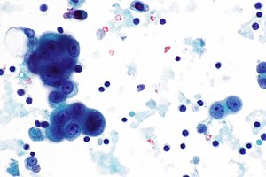
Mesothelioma In Cytology - Proceedings Of The American Society Of Cytopathology Companion Session At The 2019 United States And Canadian Academy Of Pathology Annual Meeting Part 2 Effusion Cytology With Focus On Theranostics And Diagnosis Of : Cytology description adenocarcinoma appears as a distinct population from background mesothelial cells, while mesothelioma appears as a uniform population adenocarcinoma is the likeliest lung cancer cell type to generate a malignant pleural effusion and it is also associated with the highest cytological yield (ann transl med 2019;7:352)
Mesothelioma In Cytology - Proceedings Of The American Society Of Cytopathology Companion Session At The 2019 United States And Canadian Academy Of Pathology Annual Meeting Part 2 Effusion Cytology With Focus On Theranostics And Diagnosis Of : Cytology description adenocarcinoma appears as a distinct population from background mesothelial cells, while mesothelioma appears as a uniform population adenocarcinoma is the likeliest lung cancer cell type to generate a malignant pleural effusion and it is also associated with the highest cytological yield (ann transl med 2019;7:352)
Mesothelioma In Cytology - Proceedings Of The American Society Of Cytopathology Companion Session At The 2019 United States And Canadian Academy Of Pathology Annual Meeting Part 2 Effusion Cytology With Focus On Theranostics And Diagnosis Of : Cytology description adenocarcinoma appears as a distinct population from background mesothelial cells, while mesothelioma appears as a uniform population adenocarcinoma is the likeliest lung cancer cell type to generate a malignant pleural effusion and it is also associated with the highest cytological yield (ann transl med 2019;7:352). A rare case of primary pulmonary ss in an asymptomatic. Find updated content daily for how do you get mesothelioma. These results suggest that cytokeratin 5/6 is neither a sensitive nor specific stain for the diagnosis of mesothelioma in cytology material. Unlike imaging tests, cytology is a pathological means to diagnosing malignant mesothelioma in patients, consisting of a fluid biopsy of abnormalities previously discovered. At the study institution (northwestern university), a primary diagnosis of mm is made on fluid cytology specimens.
This is yet another reason mesothelioma is difficult to diagnose. The cytologic characteristics of mesothelioma are widely variable. While the histological determination will focus more on the cell type, the cytology research will dive deeper into how these cells are forming, interacting and spreading in the body. Pleural biopsy or cytology were rarely contributory. Once sample cells are collected from a patient, they are sent to a laboratory for review.
Mesothelial Cytopathology Libre Pathology from librepathology.org Once cytology is performed, the cells from the sample are examined to arrive at a diagnosis. The article deals with cytopathology specimens from spaces lined with mesothelium, i.e. By forgoing a tissue biopsy, there is less risk of morbidity of the patient. Purpose of review advances in the pathology and genetics of malignant pleural mesothelioma (mpm) have impacted upon cytology diagnosis. Mesothelioma cytology or mesothelioma cytopathology is the study of cells for the presence of mesothelioma. Recent findings the diagnostic accuracy of. Mesothelioma diagnosis in effusion cytology/paintal et al cancer cytopathology december 2013 705. Mesothelioma cytology, or mesothelioma cytopathology, is the study of cells for the presence of mesothelioma.
In biphasic mesothelioma tumors, there are both types of cells.
Mesothelioma diagnosis in effusion cytology/paintal et al cancer cytopathology december 2013 705. Types of mesothelioma cancer cells In rare cases, mesothelioma tumors can grow in the linings of the heart. Find updated content daily for how do you get mesothelioma. The cytology of malignant mesothelioma seen in 12 patients associated with the mining of crocidolite asbestos in western australia is described. An introduction to cytopathology is in the cytopathology article. Cytopathology is the inspection of cells for diseases. The cytologic characteristics of mesothelioma are widely variable. However, there is still a small risk of tumor cell seeding (microscopic spreading of cancer due to cells moving during a biopsy) during cytologic procedures. Cytology may be the first step that a doctor takes to gather information about mesothelioma because the procedures are minimally invasive. It deals with pericardial fluid, peritoneal fluid and pleural fluid. Mesothelial cytopathology is a large part of cytopathology. These results suggest that cytokeratin 5/6 is neither a sensitive nor specific stain for the diagnosis of mesothelioma in cytology material.
Pleural effusions are among the first clinical manifestations of malignant pleural mesothelioma (mpm) and often constitute the only available material for diagnosis. Distinguishing between the two is very difficult. While the histological determination will focus more on the cell type, the cytology research will dive deeper into how these cells are forming, interacting and spreading in the body. To distinguish mesothelioma from adenocarcinoma or reactive mesothelial cells is difficult, and consequently the diagnostic accuracy by cytology is not high. At the study institution (northwestern university), a primary diagnosis of mm is made on fluid cytology specimens.
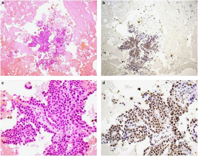
Loss Of Expression Of Bap1 Is A Useful Adjunct Which Strongly Supports The Diagnosis Of Mesothelioma In Effusion Cytology Modern Pathology from media.springernature.com Mesothelial cytopathology is a large part of cytopathology. But the science of cytology is currently not considered as accurate as a histological sample to diagnose mesothelioma. Once sample cells are collected from a patient, they are sent to a laboratory for review. At the study institution (northwestern university), a primary diagnosis of mm is made on fluid cytology specimens. The authors also evaluated their own institution's experience. Types of mesothelioma cancer cells Über 7 millionen englischsprachige bücher. The mesothelial cells have central round nuclei with a moderate amount of light purple cytoplasm and a corona or fringe to the cytoplasmic borders.
In an effort to estimate the practice at other institutions, a survey was disseminated regarding cytologic diagnosis of mm.
Once sample cells are collected from a patient, they are sent to a laboratory for review. The cytology of malignant mesothelioma seen in 12 patients associated with the mining of crocidolite asbestos in western australia is described. Cytology is the study of cells coming from either a fluid specimen or very small tissue. Fish, in contrast, is rapidly emerging as an extremely useful confirmatory tool in the diagnosis of mm. American society of clinical oncology clinical practice guideline. Find updated content daily for how do you get mesothelioma. Cytology description adenocarcinoma appears as a distinct population from background mesothelial cells, while mesothelioma appears as a uniform population adenocarcinoma is the likeliest lung cancer cell type to generate a malignant pleural effusion and it is also associated with the highest cytological yield (ann transl med 2019;7:352) By forgoing a tissue biopsy, there is less risk of morbidity of the patient. The broad term cytology refers to the study of cell structure and how cells function. Mesothelioma cytology is a useful tool that enables analysis without necessitating a tissue biopsy. In an effort to estimate the practice at other institutions, a survey was disseminated regarding cytologic diagnosis of mm. Although an mpm diagnosis can be reliable on cytology, the reported sensitivity is low (30% to 75%). A rare case of primary pulmonary ss in an asymptomatic.
Case contributed by dr annabelle mahar, sydney, australia. Mesothelioma cytology, or mesothelioma cytopathology, is the study of cells for the presence of mesothelioma. To distinguish mesothelioma from adenocarcinoma or reactive mesothelial cells is difficult, and consequently the diagnostic accuracy by cytology is not high. By forgoing a tissue biopsy, there is less risk of morbidity of the patient. In an effort to estimate the practice at other institutions, a survey was disseminated regarding cytologic diagnosis of mm.
Malignant Mesothelioma Cytology from media.curofy.com Mesothelial cytopathology is a large part of cytopathology. However, there is still a small risk of tumor cell seeding (microscopic spreading of cancer due to cells moving during a biopsy) during cytologic procedures. The authors also evaluated their own institution's experience. Particularly, it can be hard to discriminate epithelioid mpm, the most common histotype, from reactive mesothelial hyperplasia (mh). At the study institution (northwestern university), a primary diagnosis of mm is made on fluid cytology specimens. Malignant mesothelioma is a cancer caused by asbestos fibers. Cells that resemble those found in a condition called fibrous pleuritic. These results suggest that cytokeratin 5/6 is neither a sensitive nor specific stain for the diagnosis of mesothelioma in cytology material.
Recent findings the diagnostic accuracy of.
Mesothelioma cytology, or mesothelioma cytopathology, is the study of cells for the presence of mesothelioma. Background the diagnosis of malignant mesothelioma (mm) in effusion specimens is controversial. In rare cases, mesothelioma tumors can grow in the linings of the heart. It deals with pericardial fluid, peritoneal fluid and pleural fluid. Fish, in contrast, is rapidly emerging as an extremely useful confirmatory tool in the diagnosis of mm. The diagnosis of malignant mesothelioma (mm) in effusion specimens is controversial. Numerous mesothelial cells are seen in this pleural fluid from a dog with a transudative effusion (with concurrent diapedesis of red blood cells or hemorrhage). Cells that resemble those found in a condition called fibrous pleuritic. To distinguish mesothelioma from adenocarcinoma or reactive mesothelial cells is difficult, and consequently the diagnostic accuracy by cytology is not high. Pleural effusions are among the first clinical manifestations of malignant pleural mesothelioma (mpm) and often constitute the only available material for diagnosis. Follow the guidelines, common sense, clinical/imaging findings, and remember, behind every glass slide there is a human being and soon… Cytology can identify the presence of an individual malignant cancerous cell, but cytology cannot differentiate between a malignant tumor, a. A look at how the cancer will advance.

0 comments